Atomic-scale study of defects in van der Waals heterostructures
Atomic-scale study of defects in van der Waals heterostructures
Research questions:
Of the large number of currently known 2D materials, TMDs offer arguable the largest variety of properties. Beyond the individual materials, the application potential of 2D materials can be further increased through their combination in heterostructures [1]. However, many of the TMDs have been reported to contain a large number of defects. It is widely believed that these defects are responsible for interesting optical properties of TMDCs [2]. In this project, we create heterostructures of graphene and inorganic 2D materials [3] and study both their intrinsic defects as well as introduce defects via ion and electron irradiation.
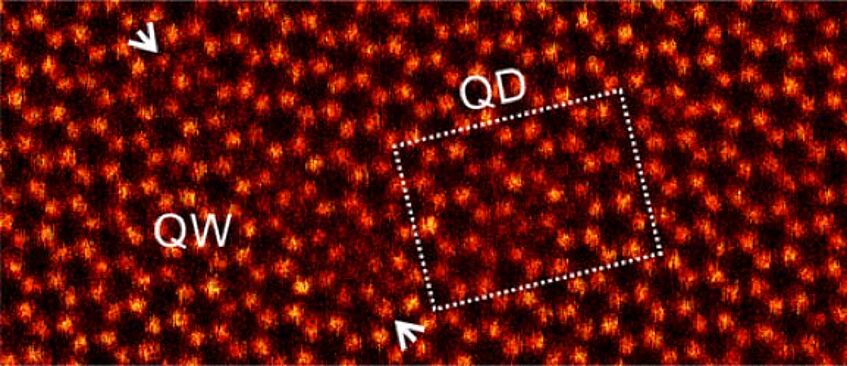
Intrinsic defects in MoTe2. [4]
Methods:
The atomic structure of the created structures will be imaged directly using transmission electron microscopy (TEM) using both high resolution TEM (FEI Titan 80–300) that allows phase contrast imaging, energy dispersive X-ray spectroscopy and electron diffraction and scanning TEM (STEM) (Nion UltraSTEM 100) that allows Z-contrast imaging and atomic-scale electron energy loss spectroscopy [5]. Defects will be created both in situ inside the microscopes using the energetic imaging electrons as well as in the manipulation setup that shares the ultra high vacuum system with the Nion microscope allowing defect creation and imaging without breaking the vacuum and thus exposing the structures to air. These studies can be complemented by the study of chemical stability of the structures by exposing them to different molecular atmospheres in the Nion microscope in situ [6].
Time frame:
Months 1-6: preparation of samples using mechanical exfoliation and commercial graphene samples; months 7-18: study of intrinsic defects; months 19-30: study of defects created in situ in the microscope; months 31-42: study of defects created in the manipulation setup and chemical reactivity; months 43-48: writing of papers and thesis.
Participating DCAFM-faculty:
J. Kotakoski (PI), C. Dellago (neural network simulation of defects), C. Franchini (ab initio training set for machine learning), T. Pichler (optical spectroscopy).
[1] Novoselov, K. S. et al., Science 353, aac9439 (2016), DOI: 10.1126/science.aac9439.
[2] Hong, J., Jin, C., Yuan, J.& Zhang, Z. Adv. Mater. 29, 1606434 (2017), DOI:10.1002/adma.201606434.
[3] Argentero, G. et al., Nano Lett. 17, 1409–1416 (2017), DOI: 10.1021/acs.nanolett.6b04360.
[4] Elibol, K. et al., Chem. Mater. 30, 1230–1238 (2018), DOI: 10.1021/acs.chemmater.7b03760.
[5] Susi, T. et al., 2D Mater. 4, 021013 (2017), DOI: 10.1088/2053-1583/aa5e78.
[6] Leuthner, G. T. et al., Ultramicroscopy, in press (2019), DOI: 10.1016/j.ultramic.2019.02.002.